http://www.ncbi.nlm.nih.gov/pmc/articles/PMC2807988/
The biological perception of light is mediated by a collection of photoreceptors that couple light absorption to specific signaling cascades. One influential set is the phytochromes, a superfamily of dimeric chromoproteins that absorb light via a bound bilin (or linear tetrapyrrole) chromophore
1,
2,
8. The bilin is buried within an N-terminal cGMP phosphodiesterase/adenyl cyclase/FhlA (GAF) domain whose contacts with the chromophore generate much of the unique photochromic behavior of Phys. Typically, the GAF domain is preceded by a Per/Arndt/Sim (PAS) domain and followed by a Phy-associated (PHY) domain and an output module, which often includes a histidine kinase domain that initiates a two-component phosphorelay. By photointerconversion between Pr and Pfr, phytochromes act as light-regulated switches for measuring the fluence, direction, duration and color of the ambient light environment
8.
Despite intensive study, we know little about how phytochromes acquire their unique photochromic behavior and how Pfr then initiates signal transmission. Recently, we and others provided important insights by determining the structure of the bilin-binding photosensory domain as Pr
3-
7. These models showed that the bilin is cradled within the GAF domain crevice, revealed a figure-of-eight knot that connects the PAS and GAF domains, identified a dimerization contact between adjacent GAF domains in the homodimer, and discovered a hairpin projection from the PHY domain that helps seal the chromophore pocket from the solvent. Unfortunately, these models have not fully illuminated how Pfr is generated. A long held notion is that the initial photochemistry involves a
Z to
E isomerization of the C15=C16 methine bridge which concomitantly rotates the D pyrrole ring
9-
13. Specific protein conformational changes have also been proposed from the structural analyses of an unusual phytochrome variant that prefers Pfr as the ground state, but whether these movements pertain to canonical phytochromes remains speculative
7,
14.
To better understand photoconversion, we used NMR spectroscopy to generate companion high resolution Pr and Pfr structures of the GAF domain from the phytochrome
SyB-Cph1 obtained from the thermotolerant cyanobacterium
Synechococcus OSB’. This fragment efficiently assembles with its native chromophore phycocyanobilin (PCB) to generate a chromoprotein with near full Pr/Pfr photochromicity
4,
15. NMR spectra were collected without illumination with the chromoprotein as Pr and during continuous red light irradiation, which produced an equimolar mixture of Pr and Pfr. By comparing the results to our previous
SyB-Cph1(GAF) Pr structure
4, we generated a highly refined solution structure of Pfr (Protein Data Bank (PDB) code 2KLI) and an improved solution structure of Pr (PDB code 2KOI) with structured backbone root mean square deviations of 0.44 Å and 0.30 Å, respectively.
The backbone conformation of the
SyB-Cph1 GAF domain as Pfr is similar to that as Pr, indicating that the overall shape of this domain does not change dramatically during photoconversion (
Fig. 1). However, photoinduced movements were obvious for the bilin and a number of amino acid side chains. In contrast to our previous report
4, the refined Pr structure showed that the PCB A pyrrole ring is nearly perpendicular to the B and C rings, with the A-ring carbonyl now pointing away from the thioether linkage to Cys138 (
Fig. 2a,b). Upon photoconversion to Pfr, the orientations of the B, C and D rings are unchanged. Instead, we found in the ensemble of Pfr conformers that the A ring becomes nearly co-planar with the B and C rings, implying a ~90° rotation around the C4=C5 bridge during photoconversion (
Fig. 2b). The thioether linkage to PCB is also contorted, which is supported by the fact that the Cys138 β carbon displays the largest chemical shift change during photoconversion (-4.6 ppm (
Suppl. Fig. 1)). Most NMR signals from PCB exhibited considerable broadening in Pfr, suggesting increased mobility relative to the more rigid Pr state (
Fig. 2c,d and
Suppl. Fig. 2).

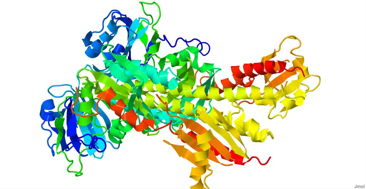
Figure 1
Three-dimensional overlay of SyB-Cph1(GAF) Pr and Pfr solution structures

Figure 2
Rotation of the A ring of the PCB chromophore during Pr to Pfr photoconversion
Although prior studies proposed that the D ring rotates during phototransformation
9-
11,
13, our NMR analyses of
SyB-Cph1(GAF) failed to detect significant chemical shift changes for this ring during photoconversion. For example, various NMR spectra for the D-ring C171 and C182 methyls, amide, the pyrrole nitrogen, and C18 failed to detect Pfr signals distinct from Pr, nor did the immediate neighboring C131 methyl of the C ring, whereas differences in and around the environment of the A ring were obvious (
Fig. 2c,d,
Suppl. Fig. 2, and ref.
15).
Rotation of the A ring of
SyB-Cph1(GAF) is accompanied by conformational changes of several amino acids proximal to PCB, including Asp86, Tyr142, Phe82, Tyr54, His139, His169, Arg101 and Val100. Previous structural studies of Pr showed that the Nδ1 nitrogen of His139 contributes to a complex hydrogen bond network, involving the A-C ring nitrogens and a centrally positioned pyrrole water which together participate in the protonation cycle of the bilin during photoconversion, whereas the Nψ1 nitrogen of His169 hydrogen bonds with the C19 carbonyl oxygen to stabilize the D ring
4-
6,
16,
17. In Pfr, both these interactions are disrupted; the imidazole rings of His139 and His169 are rotated away from the pyrrole water and the D ring, respectively (
Fig 3a-c). The position of His169 in Pfr is stabilized by displacement of strand β6 toward strand β1, leading to the formation of a new set of hydrogen bonds involving His170 with Tyr176 and Thr48 (
Suppl. Fig. 3). Collectively, these changes likely alter the environment of the pyrrole water and thus the bilin photocycle
16-
19, a possibility supported by our observations that the Pfr forms of Tyr176-Phe, His169-Ala, and Thr48-Ala mutants thermally revert more rapidly back to Pr (
Suppl. Fig. 4).

Figure 3
Light-driven conformational changes for amino acids surrounding the chromophore
A second set of rearrangements during photoconversion involves Phe82, Tyr54, Asp86, and Tyr142 near the A and D rings of PCB (Figs.
3a-c and
4a,b). The Phe82 aromatic ring rotates ~30° to assume a parallel displaced orientation relative to the PCB D ring that could enable hydrophobic π stacking interactions (
Fig. 4e,f). Movement of Phe82 eliminates a hydrogen bond between its main chain nitrogen and the hydroxyl of Tyr54, a conserved residue that helps avoid non-productive fluorescence of some Phys during photoexcitation
15,
16,
20. Mutant analysis shows that both Phe82 and Tyr54 are required for Pfr formation and stability (
Suppl. Fig. 4). Rotation of the A-ring nitrogen to its position in Pfr is stabilized by a new hydrogen bond with the main chain oxygen of Asp86. Subsequent motion of the Asp86 side chain then leads to a new hydrogen bond network with the hydroxyl of Tyr142 and the D-ring carbonyl, the importance of which is confirmed by the aberrant photochemistry of a Tyr142-Phe mutant and several Asp86 substitutions (
Suppl. Fig. 4 and ref.
15). Collectively, these movements help stabilize the D ring (in addition to its contact with Lys52), and decrease the solvent accessibility of the Pfr chromophore (
Fig 4c,d). Given that the carboxylate group of Asp86 is predicted to form a double salt bridge with a conserved arginine located in the PHY domain hairpin
3,
7, movement of Asp86 likely affects this contact as well.

Figure 4
Conformational rearrangement of Asp86, Tyr142 and Phe82 during Pr to Pfr photoconversion
Perhaps the most dramatic change in the
SyB-Cph1(GAF) domain during photoconversion involves movement of Arg101 (
Fig. 3d-f). In Pr, Arg101 forms a double salt bridge with the carboxylate of the B-ring propionate, but in Pfr, strand β4 is disrupted, and Arg101 and Val100 swivel approximately 180° to encourage a salt bridge between Arg101 and Glu185 in helix α5 (
Fig. 3d-f and
Suppl. Fig. 5). Concomitant with this rotation is a 2.6 Å displacement of helix α2 toward the B-ring propionate, thus allowing Phe95 to fill the void left by Arg101. Previous mutagenic analyses of Arg101 and a comparable Arg in Arabidopsis PhyB revealed a critical role for this residue in Pfr stability and signaling
4,
22, whereas mutagenic studies of both Gln185 and Arg101 support the importance of their contact in Pfr (
Suppl. Fig. 4). For example, the Gln185-Ala and Gln185-Glu mutants of
SyB-Cph1 thermally revert from Pfr to Pr slower than wild type, whereas the Gln185-Arg and Arg101-Ala mutants revert much faster, with the Gln185-Arg mutant also displaying aberrant absorption spectra. Collectively, the new Pfr contact between Arg101 and Gln185 appears to adjust the position and/or flexibility of helix α5, as detected by notable chemical shift changes for several neighboring residues (
e.g. Val184) and the unusual absence of NMR signals in Pfr from helix α5 (
e.g., Gln178, Glu179, Glu180, Leu181, and Gln185), which participates in Phy dimerization
6,
7.
Taken together, the structural differences between Pr and Pfr in
SyB-Cph1(GAF) combined with the photochemical importance of a number of key conserved residues (
Suppl. Fig. 6) offer a possible model for phytochrome photoconversion. An unexpected feature is the substantial rotation of the A ring, presumably driven by a C4=C5 isomerization, and a contortion of its thioether linkage to the protein instead of the former proposal that the D ring rotates
9-
13. Either isomerization or relaxation of the strained C4=C5 bridge during Pfr formation could account for the red-shifted absorption spectrum of Pfr by increasing the coupling of the π-conjugation system. Further red-shifting by π-stacking interactions with aromatic residues neighboring the D ring could help explain the Pfr/Pr chemical shift differences reported previously for the D ring
23. Rotation of the A ring is also supported by photochemical studies with sterically locked bilins
24,
25 and by prior NMR spectra of phytochrome fragments also containing the PAS and PHY domains
26,
27, which strongly suggest that our results with
SyB-Cph1(GAF) are not unique to this phytochrome nor artifactually generated by analysis of just the GAF domain. We note that the x-ray crystallographic structures of several Phys as Pr
3-
5,
7,
28 have modeled with a more coplanar configuration for the A ring relative to the B and C rings than seen here for
SyB-Cph1 in solution. These differences could reflect subtle variations among Phys, inherent differences in the environment of the chromophore in crystals versus in solution, and/or radiation-induced damage of the Pr bilin during x-ray data collection that could relax the strain of a non-planar A ring
5,
28.
We propose that, subsequent to rotation of the A ring, a series of reversible conformational movements occur within the bilin-binding pocket that support the deprotonation/protonation cycle of the bilin, stabilize the Pfr form, and finally adjust several contact sites on the surface of the GAF domain. In particular, movement of the Asp86 and Tyr142 pair could affect the non-covalent interaction of the PHY domain with the GAF domain through its hairpin projection, which could then reorient by a hinge mechanism these domains relative to each other. The swivel of Arg101 to contact Gln185 concomitantly reorients and/or destabilizes helix α5. Given the role of helix α5 in helping sister phytochromes dimerize and in covalently connecting the GAF and PHY domains
3,
6,
7, even a subtle movement/unfolding of this helix might have profound consequences on intermolecular GAF/GAF dimerization and intramolecular GAF/PHY contacts.
Taken together, it is conceivable that such light-induced rearrangements then initiate a cascade of events within the phytochrome dimer that reorient the C-terminal output modules relative to each other and to the photosensory modules. For phytochromes bearing histidine kinase output modules, such light-driven rearrangements could then alter autophosphorylation
in trans across the phytochrome dimer. In this manner, phytochromes may resemble the phototropin family of photoreceptors, which couples flavin photochemistry to selective destabilization of a helical contact adjacent to the photosensory domain and finally to activation of the appended output kinase
29. Because the PYP family of photoreceptors may work by a similar light-triggered conformational switch
30, our model for phytochromes provides further support for the notion that light-induced conformational changes are fundamental for photoactivated signaling.